Animal Cell Facts Biography
Source:- Google.com.pk
Cells
All living organisms are comprised of one or more cells. Most animal cells range in size between 10 and 100 micrometers and have several key elements.
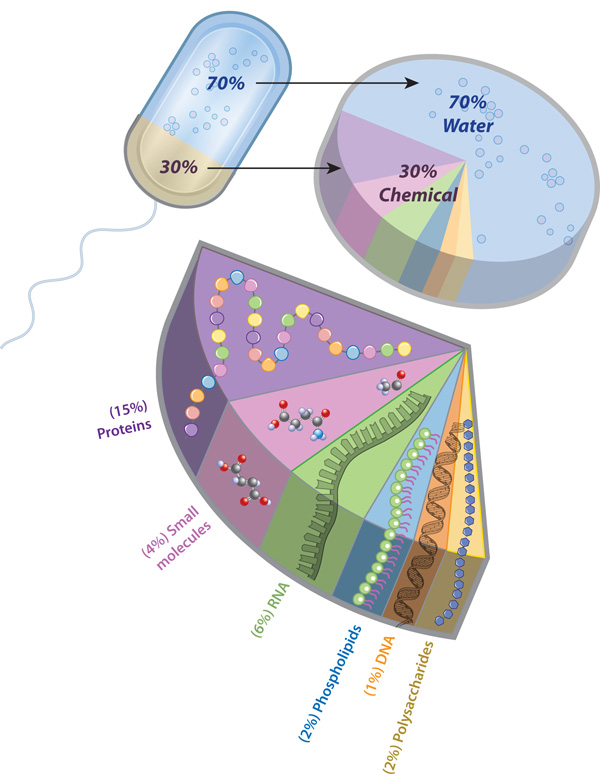
The outer layer of a cell, the cell membrane, consists of a phospholipid bilayer, which serves primarily as a barrier from the external environment, and integral proteins which function mainly to regulate transport. The cell membrane creates an enclosed space in which the chemical processes, or metabolism, of the cell can occur. It also serves as a gatekeeper for the cell, carefully regulating what passes in and out. For example, nutrients pass in, and metabolic waste passes out.
The inside of an animal cell contains several organelles , specialized structures that perform specific functions for the cell. These organelles are suspended in a thick aqueous solution, the cytoplasm. The coordinated activity of the organelles is required for the survival of the cell. Many organelles are enclosed by membranes that generate separate compartments within the cytoplasm. One of the most important compartments is the nucleus. The nucleus contains genetic material, which acts as a blue print for the production of the proteins that perform most cellular functions. Protein synthesis is performed by ribosomes and takes place in the cytoplasm. Ribosomes attach to the endoplasmic reticulum (ER) during synthesis of those proteins that are to be exported, incorporated into the membrane, or placed into organelles. These proteins are then shipped from the ER to another compartment, the Golgi apparatus, where they are modified and then shipped to their final destinations. In recycling compartments, known as the lysosomes, old proteins and other molecules are broken down so that their components can be reused. Diverse chemical processes (e.g. the synthesis of the gas Nitric Oxide that functions as an important signal between cells) that produce toxic molecules (peroxides) as side products, take place in specialized chemical compartments of the cell which are called peroxisomes. As a result, the peroxides can be broken down safely within the peroxisomes without harming the cell.
The transport of cargo between individual compartments through the cytoplasm is the job of transport vesicles, tiny membrane-enclosed compartments that contain the cargo and transport it through the cytoplasm. Every cell produces many transport vesicles, and each type is specialized for a distinct shipping route within the cell, for a kind of cargo, or even for the storage of substances (e.g., neurotransmitters for communication between cells).
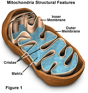
The energy for all the chemical processes in the cell is generated in compartments called mitochondria , which can be considered the cell's powerhouses. Mitochondria produce ATP, the energy source of the cell, using sugars and oxygen in a process called oxidative phosphorylation.
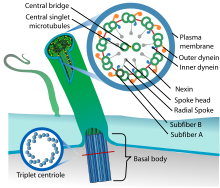
The shape of cells is maintained by a cytoskeleton, or cell skeleton, made of three membrane-free organelles—microtubules, actin filaments, and intermediate filaments. Together these organelles form a network of molecular cables and struts that stabilizes the cell shape.
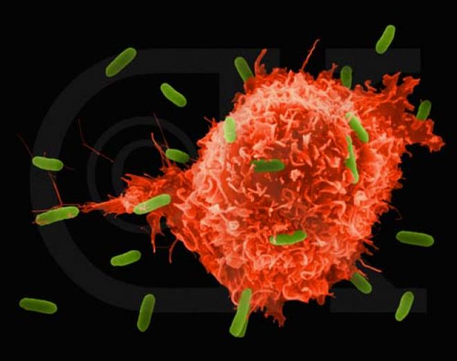
In contrast to plant cells, which are further stabilized by a cell wall that surrounds the outer cell membrane, animal cells are stabilized by a cytoskeleton and an extracellular matrix made mostly of glycoproteins . However, because they are less rigid, animal cells can change their shape more easily and even use these shape changes to move. Animal cells such as the infection-fighting white blood cells can be sophisticated "crawlers." In the absence of a rigid cell wall, they assemble and disassemble parts of their cytoskeleton in such a way that specific shape changes leading to cell movement will occur. Microfilaments are also responsible for the movement of specific organelles within the cell, and microfilaments and microtubules together are essential for cell division. While microtubules ensure the distribution of duplicated chromosomes to the two daughter cells, the microfilaments will finish the separation of the original cell by pinching in the outer cell membrane.
Whereas in single-celled organisms all life functions are performed by a single cell, in multicellular organisms, such as animals, division of labor and specialization among cells occurs. For example, humans have about 200 different cell types that differ in structure and in function. In all but the simplest animals, the sponges, specialized cells that have a similar structure and function are arranged together into tissues. Although there are many different types of animal cells, scientists group them all into only four general tissue types—epithelial tissue, muscle tissue, nervous tissue, and connective tissue . The cells in a tissue may be held together by the extracellular matrix that makes the cells sticky or ties them together.
In all animals except sponges and jellyfishes, different tissue types may form a functioning unit called an organ. Organs may also be part of an organ system, such as the digestive system and reproductive system, where several organs function together. Each organ system has a different function, but just like the organelles within an individual cell, the function of each organ system must be regulated and coordinated to ensure the survival of the whole animal.
see also Cell Division; Homeostasis.
Kathrin F. Stanger-Hall
Bibliography
Barnes-Svarney, Patricia. New York Public Library Desk Reference. New York: Macmillan USA, 1998.
Ingber, D. E. "The Architecture of Life." Scientific American 278 (1998):48-57.
Rhoades, Rodney, and Richard Pflanzer. Human Physiology, 3rd ed. Fort Worth, TX: Saunders College Publishing, 1996.
Rothman, James E. "Budding Vesicles in Living Cells." Scientific American 274 (1996):70-75.
Cite this article
Pick a style below, and copy the text for your bibliography


All living organisms are comprised of one or more cells. Most animal cells range in size between 10 and 100 micrometers and have several key elements.
The outer layer of a cell, the cell membrane, consists of a phospholipid bilayer, which serves primarily as a barrier from the external environment, and integral proteins which function mainly to regulate transport. The cell membrane creates an enclosed space in which the chemical processes, or metabolism, of the cell can occur. It also serves as a gatekeeper for the cell, carefully regulating what passes in and out. For example, nutrients pass in, and metabolic waste passes out.
The inside of an animal cell contains several organelles , specialized structures that perform specific functions for the cell. These organelles are suspended in a thick aqueous solution, the cytoplasm. The coordinated activity of the organelles is required for the survival of the cell. Many organelles are enclosed by membranes that generate separate compartments within the cytoplasm. One of the most important compartments is the nucleus. The nucleus contains genetic material, which acts as a blue print for the production of the proteins that perform most cellular functions. Protein synthesis is performed by ribosomes and takes place in the cytoplasm. Ribosomes attach to the endoplasmic reticulum (ER) during synthesis of those proteins that are to be exported, incorporated into the membrane, or placed into organelles. These proteins are then shipped from the ER to another compartment, the Golgi apparatus, where they are modified and then shipped to their final destinations. In recycling compartments, known as the lysosomes, old proteins and other molecules are broken down so that their components can be reused. Diverse chemical processes (e.g. the synthesis of the gas Nitric Oxide that functions as an important signal between cells) that produce toxic molecules (peroxides) as side products, take place in specialized chemical compartments of the cell which are called peroxisomes. As a result, the peroxides can be broken down safely within the peroxisomes without harming the cell.
The transport of cargo between individual compartments through the cytoplasm is the job of transport vesicles, tiny membrane-enclosed compartments that contain the cargo and transport it through the cytoplasm. Every cell produces many transport vesicles, and each type is specialized for a distinct shipping route within the cell, for a kind of cargo, or even for the storage of substances (e.g., neurotransmitters for communication between cells).
The energy for all the chemical processes in the cell is generated in compartments called mitochondria , which can be considered the cell's powerhouses. Mitochondria produce ATP, the energy source of the cell, using sugars and oxygen in a process called oxidative phosphorylation.
The shape of cells is maintained by a cytoskeleton, or cell skeleton, made of three membrane-free organelles—microtubules, actin filaments, and intermediate filaments. Together these organelles form a network of molecular cables and struts that stabilizes the cell shape.
In contrast to plant cells, which are further stabilized by a cell wall that surrounds the outer cell membrane, animal cells are stabilized by a cytoskeleton and an extracellular matrix made mostly of glycoproteins . However, because they are less rigid, animal cells can change their shape more easily and even use these shape changes to move. Animal cells such as the infection-fighting white blood cells can be sophisticated "crawlers." In the absence of a rigid cell wall, they assemble and disassemble parts of their cytoskeleton in such a way that specific shape changes leading to cell movement will occur. Microfilaments are also responsible for the movement of specific organelles within the cell, and microfilaments and microtubules together are essential for cell division. While microtubules ensure the distribution of duplicated chromosomes to the two daughter cells, the microfilaments will finish the separation of the original cell by pinching in the outer cell membrane.
Whereas in single-celled organisms all life functions are performed by a single cell, in multicellular organisms, such as animals, division of labor and specialization among cells occurs. For example, humans have about 200 different cell types that differ in structure and in function. In all but the simplest animals, the sponges, specialized cells that have a similar structure and function are arranged together into tissues. Although there are many different types of animal cells, scientists group them all into only four general tissue types—epithelial tissue, muscle tissue, nervous tissue, and connective tissue . The cells in a tissue may be held together by the extracellular matrix that makes the cells sticky or ties them together.
In all animals except sponges and jellyfishes, different tissue types may form a functioning unit called an organ. Organs may also be part of an organ system, such as the digestive system and reproductive system, where several organs function together. Each organ system has a different function, but just like the organelles within an individual cell, the function of each organ system must be regulated and coordinated to ensure the survival of the whole animal.
see also Cell Division; Homeostasis.
Kathrin F. Stanger-Hall
Bibliography
Barnes-Svarney, Patricia. New York Public Library Desk Reference. New York: Macmillan USA, 1998.
Ingber, D. E. "The Architecture of Life." Scientific American 278 (1998):48-57.
Rhoades, Rodney, and Richard Pflanzer. Human Physiology, 3rd ed. Fort Worth, TX: Saunders College Publishing, 1996.
Rothman, James E. "Budding Vesicles in Living Cells." Scientific American 274 (1996):70-75.
Cite this article
Pick a style below, and copy the text for your bibliography
Animal Cell Facts Animal Cell Model Diagram Project Parts Structure Labeled Coloring and Plant Cell Organelles Cake
Animal Cell Facts Animal Cell Model Diagram Project Parts Structure Labeled Coloring and Plant Cell Organelles Cake
Animal Cell Facts Animal Cell Model Diagram Project Parts Structure Labeled Coloring and Plant Cell Organelles Cake
Animal Cell Facts Animal Cell Model Diagram Project Parts Structure Labeled Coloring and Plant Cell Organelles Cake
Animal Cell Facts Animal Cell Model Diagram Project Parts Structure Labeled Coloring and Plant Cell Organelles Cake
Animal Cell Facts Animal Cell Model Diagram Project Parts Structure Labeled Coloring and Plant Cell Organelles Cake
Animal Cell Facts Animal Cell Model Diagram Project Parts Structure Labeled Coloring and Plant Cell Organelles Cake
Animal Cell Facts Animal Cell Model Diagram Project Parts Structure Labeled Coloring and Plant Cell Organelles Cake
Animal Cell Facts Animal Cell Model Diagram Project Parts Structure Labeled Coloring and Plant Cell Organelles Cake
Animal Cell Facts Animal Cell Model Diagram Project Parts Structure Labeled Coloring and Plant Cell Organelles Cake
Animal Cell Facts Animal Cell Model Diagram Project Parts Structure Labeled Coloring and Plant Cell Organelles Cake
No comments:
Post a Comment