Do Animal Cells Have Vacuoles Biography
Source:- Google.com.pk
Compare the surface to volume ratio (surface:volume) of a cube that is 1 cm X 1 cm X 1 cm with that of a cube that is 10 cm X 10 cm X 10 cm.
Smaller cube (1 cm X 1 cm X 1 cm)
The surface area of one side = 1 cm X 1 cm = 1 square cm (or 1 cm2).
There are 6 sides, so the total surface area = 6 X cm2 = 6 cm2.
Volume = 1 cm X 1 cm X 1 cm = 1 cubic cm (or 1 cm3)
Surface:Volume = 6 cm2/1 cm3 = 6 cm2/cm3 (or 6 square cm of surface area for each cubic cm of volume)
Larger cube (10 cm X 10 cm X 10 cm)
The surface area of one side = 10 cm X 10 cm = 100 square cm (or 100 cm2).
There are 6 sides, so the total surface area = 600 X cm2 = 600 cm2.
Volume = 10 cm X 10 cm X 10 cm = 1000 cubic cm (or 1000 cm3)
Surface:Volume = 600 cm2/1000 cm3 = 0.6 cm2/cm3 (or 0.6 square cm of surface area for each cubic cm of volume).
The larger cube has more surface area and more volume but less surface area for each cubic centimeter of volume.
Nonmathematical Example
A nonmathematical example may help explain why the smaller objects have more surface for each unit of volume.
Large cube and eight smaller cubes
Imagine that a knife is used to cut the large cube three times to produce the eight smaller cubes shown above. In order to produce the smaller cubes, new surfaces were created when the knife cut the cube. The total surface area of the eight smaller cubes is greater even though the total volume has not changed.
Cells
For any given geometric object (cubes, spheres, etc.), smaller objects have a greater surface to volume ratio (surface:volume) than larger objects of the same shape.
Every cell is surrounded by a plasma membrane (discussed below and in the next chapter). Most cells are very small and therefore have a high ratio of plasma membrane surface to cell volume.
Cell Theory
All organisms are composed of cells, and a cell is the smallest unit of living matter.
Cells come only from preexisting cells.
Major Kinds of Cells
Prokaryotic Cells
Bacteria and archaea are prokaryotes. Their cells are very small and very simple. They will be discussed later.
Chapter on prokaryotes
Eukaryotic cells
All other cells are eukaryotic cells. These include protists, fungi, plants, and animals.
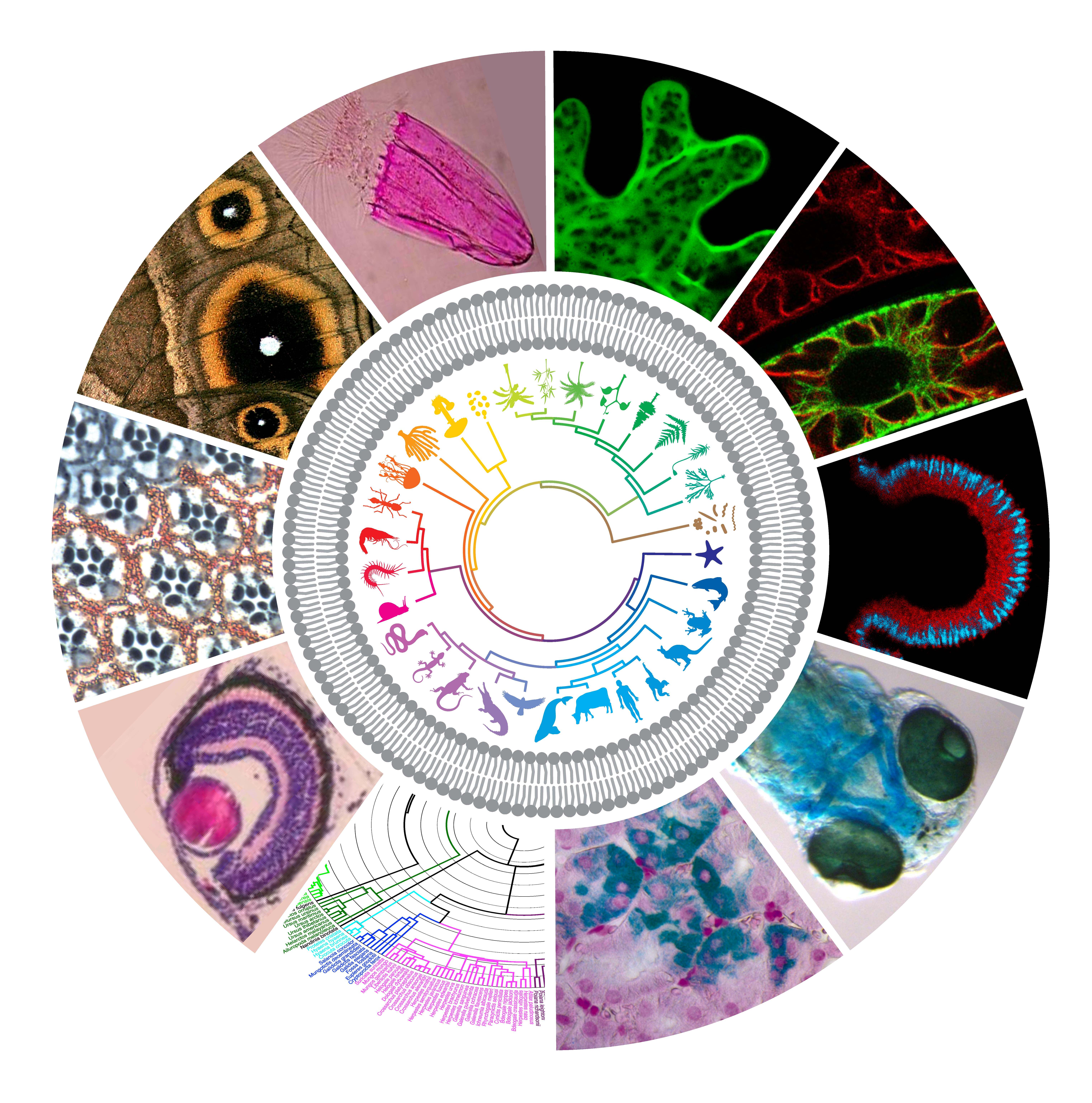
The diagram below shows evolutionary relationships between Bacteria, Archaea, and Eukarya
Eukaryotic Cells
Cells contain structures called organelles. The structure and function of the major organelles found in eukaryotic cells are described below.
Plasma membrane
All cells are surrounded by a plasma membrane. It separates the contents of the cell from its environment and regulates the passage of molecules into and out of the cell.
The membrane contains proteins that have a variety of functions. For example, some proteins are receptors which can detect the presence of certain kinds of molecules in the surrounding fluids. The function of membrane proteins will be discussed in more detail in the chapter on membranes.
An actively metabolizing cell needs a large surface area. Cells are limited in size because larger cells have a smaller surface to volume ratio.
Cells that are specialized for absorption (ex: intestinal cells) have folds in the plasma membrane called microvilli that increase the surface area.
Pseudopodia are temporary extensions of the plasma membrane used for movement or to engulf particles. Pseudopodia can be seen in the Amoeba below.
Cell Wall
The cell wall functions to support and protect the cell.
Plants have cell walls composed of cellulose; fungi have walls composed of chitin.
The cell walls of these onion skin cells can be easily seen.
Nucleus
The nuclei can be seen in the photograph of human cheek cells below.
cheek cells.jpg (21892 bytes)
The nucleus contains DNA. Recall that DNA contains instructions needed to produce proteins that control metabolism and other cell functions.
One nucleus can serve a limited amount of cytoplasm, so large cells are often multinucleate, that is, they contain more than one nucleus.
skeletal_muscle cell.jpg (85983 bytes) Teased skeletal muscle X 200
Note the many nuclei visible in the cell on the left.
The nucleus of eukaryotic cells contain a complex of DNA and proteins called chromatin. It functions to package DNA so that it fits within the nucleus and it compacts the DNA during cell division. It also plays a regulatory role in the expression of some genes. The chromatin contains several discrete pieces called chromosomes. The individual chromosomes are normally not visible but during cell division, the DNA becomes more condensed and the chromosomes become visible using light microscopy.
The material within the nucleus is referred to as the nucleoplasm.
A double membrane (nuclear envelope) surrounds the nucleus.
Nuclear pores are openings in the nuclear envelope that allow materials to pass into and out of the nucleus. The passage of RNAs, proteins, and other large molecules can be regulated by proteins associated with the pore called a pore complex.
Cytoplasm
Cytoplasm is the material enclosed by the plasma membrane, excluding the nucleus.
amoeba.jpg (56617 bytes)
Ribosomes
Ribosomes read the code in mRNA and synthesize protein accordingly.
The symbols to the left are used in the drawings of protein synthesis below.
The ribosome attaches to the mRNA.
As ribosomes move along messenger RNA (mRNA), the amino acids are added to a growing chain to form a particular protein. In these drawings, the ribosome moves from left to right.
In this drawing, the protein is nearly complete. When the ribosome reaches the end of the genetic message, it will become detached from the mRNA.
Several ribosomes may be attached to a strand of mRNA forming a unit called a polysome.
A ribosome is composed of 2 subunits. In eukaryotic cells, the subunits are synthesized in the nucleolus and move into the cytoplasm. During the process of protein synthesis, two subunits will come together along with mRNA..
Ribosomes are composed of both RNA (called ribosomal RNA or rRNA) and protein.
Some ribosomes are attached to the rough endoplasmic reticulum or the nuclear envelope and others are unattached. Ribosomes attached to the rough endoplasmic reticulum synthesize proteins directly into the space enclosed by the rough endoplasmic reticulum (the ER lumen).
Ribosomes in eukaryotes about 1/3 larger than those in prokaryotes.
Nucleolus
The nucleolus is a structure within the nucleus where the ribosomal subunits are produced.
In cells that have been stained, it appears darker than the nucleus.
Endoplasmic Reticulum
The endoplasmic reticulum is a membranous network that extends throughout the cell.
It is continuous with the nuclear envelope and the plasma membrane.
Rough Endoplasmic Reticulum
The rough appearance of rough endoplasmic reticulum is due to the presence of ribosomes on the membrane.
The rough ER functions in protein synthesis. Proteins are synthesized by ribosomes attached to the rough ER and enter the lumen (interior) of the endoplasmic reticulum while being synthesized.
The rough endoplasmic reticulum also functions in the modification of newly formed proteins. For example, some enzymes may add carbohydrate chains forming glycoproteins. Molecular chaperones are enzymes that function to fold the newly-synthesized proteins into their proper shape.
Transport vesicles are small sacs that pinch off of the endoplasmic reticulum or the Golgi apparatus (discussed below) and transport molecules to other parts of the cell.
Many of the proteins produced by the rough ER and packaged into transport vesicles are destined for secretion by the cell.
Some proteins produced by the rough ER will become packaged in organelles called lysosomes. They function in intracellular digestion.
The rough endoplasmic reticulum also functions to synthesize new membrane, including the phospholipids and embedded proteins. Transport vesicles that originate from the rough endoplasmic reticulum fuse with other membranes in the cell becoming part of the membrane
Do Animal Cells Have Vacuoles Animal Cell Model Diagram Project Parts Structure Labeled Coloring and Plant Cell Organelles Cake


Do Animal Cells Have Vacuoles Animal Cell Model Diagram Project Parts Structure Labeled Coloring and Plant Cell Organelles Cake


Do Animal Cells Have Vacuoles Animal Cell Model Diagram Project Parts Structure Labeled Coloring and Plant Cell Organelles Cake

Do Animal Cells Have Vacuoles Animal Cell Model Diagram Project Parts Structure Labeled Coloring and Plant Cell Organelles Cake


Do Animal Cells Have Vacuoles Animal Cell Model Diagram Project Parts Structure Labeled Coloring and Plant Cell Organelles Cake


Do Animal Cells Have Vacuoles Animal Cell Model Diagram Project Parts Structure Labeled Coloring and Plant Cell Organelles Cake


Do Animal Cells Have Vacuoles Animal Cell Model Diagram Project Parts Structure Labeled Coloring and Plant Cell Organelles Cake


Do Animal Cells Have Vacuoles Animal Cell Model Diagram Project Parts Structure Labeled Coloring and Plant Cell Organelles Cake


Do Animal Cells Have Vacuoles Animal Cell Model Diagram Project Parts Structure Labeled Coloring and Plant Cell Organelles Cake


Do Animal Cells Have Vacuoles Animal Cell Model Diagram Project Parts Structure Labeled Coloring and Plant Cell Organelles Cake


Do Animal Cells Have Vacuoles Animal Cell Model Diagram Project Parts Structure Labeled Coloring and Plant Cell Organelles Cake
No comments:
Post a Comment