Picture Of Animal Cell Biography
Source:- Google.com.pk
Epithelia are formed of cells that line the cavities in the body and also cover flat surfaces. Of the four major tissue types found in the human body (Figure 1), epithelial cells are by far the most prolific.
The diversity of epithelial cells makes for an interesting field of study. Our lab is dedicated to studying various aspects of rat alveolar type II (L2) lung under a variety of conditions in order to answer specific research questions. The following provides a general overview of of the six types of epithelial cells, how structure relates to function, and why research in this field is important and relevant.
Image from: http://www.pennmedicine.org/encyclopedia/em_PrintArticle.aspx?gcid=004012&ptid=1
Figure 1.The four major tissue types in humans.
Structure of Epithelial Tissue
Epithelial cells are bound together in sheets of tissue called epithelia. These sheets are held together through several types of interactions, including tight junctions, adherens, desmosomes, and gap junctions (Figure 2).One type of junction found only in epithelium is the tight junction, which is considered by most scientists as the closest junction in the world. Tight junctions act as the delineation between the apical (upper) and basal (lower) regions of an epithelial cell in conjunction with polarization between the two regions. Epithelium is supported on the basal side by a basement membrane called the basal lamina. Below the basal lamina lies the capillary bed, which provides epithelia with required nutrients and disposal of waste products. In addition, the nucleus in the epithelial cell is usually found closer to the basal surface than the apical surface.
Image from:http://cellbiology.med.unsw.edu.au/units/science/lecture0808.htm
Figure 2. Epithelia are apical to the basement membrane and are characterized by the presence of tight junctions.
How Do Epithelial Cells Differ From Other Cells?
Avascular
Capillaries do not reside within epithelial cell tissues
Sensory
Endings of neurons are present within epithelial cell tissues
Perceive external stimulus (i.e. tactile)
Gliding surface layer
Epithelial cells slough off and glide in order to replace dead cells
This function allows epithelial cells to maintain a closed barrier to the external environment
Transitional
Multi-layered epithelia are able to stretch
Allows the urinary bladder to distended or contracted without compromising it
Tight barrier
Desmosomes, hemidesmsomes, tight junctions
Epithelium is held together more tightly than other cells
Aids cells in withstanding mechanical stress
Different from endothelial cells
Endothelial cells line the insides of structures that aren’t exposed to the “outside”
Ex. Blood vessels
Where Are Epithelial Cells Found?
Epithelial cells line the major cavities of the body.
Epithelia form the structure of the lung, including the alveoli or air sacs where gas exhange occurs.
Cells line most organs, such as the stomach and small intestine, kidney, and pancreas. They also line the esophagus.
Cells are also found in ducts and glands, like the bile duct and sailvary glands.
Epithelia can specialize to act as sensory receptors. They form taste buds, line the nose, and are in the ear. They are also found in the eye.
Female reproductive organs are lined with ciliated epithelial cells.
The skin is made of epithelial cells. Its striated layers demonstrate the extensive morphology of epithelia.
Capillary beds are made of epithelium.
Epithelia is the first type of cell to differentiate in the embryo. This occurs during the eight-cell stage.
Functions (not present in every epithelial cell)
Boundary & Protection
Epithelial cells cover the inner and outer linings of body cavities, such as the stomach and the urinary tract (Figure 3). As the barrier between the outside world’s contaminants and the body, these cells replicate often to replace damaged or dead cells. Many layers provide better protection, meaning if one layer is lost, the underlying tissue is still protected. Tight junctions, are very difficult to alter or break and create a semi-permeable seal that few macromolecules or microbes can penetrate.
Image from: http://www.mc.vanderbilt.edu/histology/labmanual2002/labsection3/EsophagusandStomach03.htm
Figure 3. Layers of tissue in the upper esophagus. Shown at top is the stratified squamous epithelium, protecting the underlying tissue from damage due to outside environment exposure.
Sensory
Although epithelial cells are avascular, they are innervated. These nerve endings provide signals for sensory sensations such as taste, sight, and smell (Figure 4). These cells exhibit specialized structure to fulfill their function.
Image from: http://biodidac.bio.uottawa.ca/thumbnails/filedet.htm?File_name=L5-8J16&File_type=GIF
Figure 4. A rabbit tastebulb, showing the specialized taste receptor cells.
Transportation
Some epithelial cells, such as the ones found on the intestinal lining, aid in the transportation of filtered material through the use active-transport systems located on the apical side of their plasma membranes. For example, the glucose-Na+ symports located within certain domains of the plasma membrane of epithelial cells lining the intestine enable the cells to generate Na+ concentration gradients across their plasma membranes, which provides the energy needed to uptake glucose, from the lumen of the intestine. The glucose is then released into the underlying connective tissues and is transported into the blood supply through facilitated diffusion down its concentration gradient (Figure 5).
Image from: http://www.ncbi.nlm.nih.gov/bookshelf/br.fcgi?book=cooper&part=A1986. Permission pending.
Figure 5. Active transport of glucose in the epithelial cells lining the intestine.
Absorption
The ability of certain epithelial cells to use active-transport systems, as discussed above, enables them to absorb filtered material, such as glucose from the lumen of the intestine, which can then be circulated to the rest of the body. Cells are also able to endocytose other materials that are necessary for cell growth and signaling. For more information, see transcytosis.
Secretion & Lubrication
Some epithelial cells, such as the goblet cells, secrete fluids that are necessary for other processes such as digestion, protection, excretion of waste products, lubrication, reproduction, and the regulation of metabolic processes of the body. As part of its excretory role, certain epithelial cells secrete mucus, which lubricate the body cavities (i.e. peritoneum, pericardium, pleura, and tunica vaginalis) and passageways that they line. In the trachea, goblet epithelial cells secrete mucous which provides the lubrication to aid ciliated epithelial cells in sweeping bacteria and dust away from the lungs (Figure 6). In addition, type II alveolar cells excrete pulmonary surfactant, which decreases surface tension, allowing for normal lung function. Figure 6 shows an example of secretory cells in the fallopian tubes.
Image from: http://remf.dartmouth.edu/images/mammalianLungSEM/source/8.html
Figure 6. This scanning electron microscope image depicts the ciliated epithelial cells that line the trachea.
Movement
Some epithelial cells have cilia, which aid in moving substances in the lumen by creating a current via coordinated "sweeping" of the cilia (Figure 6). For instance, ciliated columnar epithelial cells are instrumental in the movement of the ovum through the Fallopian tubes to the uterus (Figure 7).
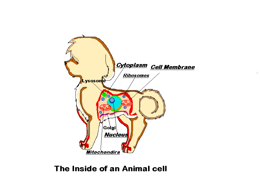
Picture Of Animal Cell Animal Cell Model Diagram Project Parts Structure Labeled Coloring and Plant Cell Organelles Cake

Picture Of Animal Cell Animal Cell Model Diagram Project Parts Structure Labeled Coloring and Plant Cell Organelles Cake
Picture Of Animal Cell Animal Cell Model Diagram Project Parts Structure Labeled Coloring and Plant Cell Organelles Cake

Picture Of Animal Cell Animal Cell Model Diagram Project Parts Structure Labeled Coloring and Plant Cell Organelles Cake

Picture Of Animal Cell Animal Cell Model Diagram Project Parts Structure Labeled Coloring and Plant Cell Organelles Cake

Picture Of Animal Cell Animal Cell Model Diagram Project Parts Structure Labeled Coloring and Plant Cell Organelles Cake

Picture Of Animal Cell Animal Cell Model Diagram Project Parts Structure Labeled Coloring and Plant Cell Organelles Cake

Picture Of Animal Cell Animal Cell Model Diagram Project Parts Structure Labeled Coloring and Plant Cell Organelles Cake

Picture Of Animal Cell Animal Cell Model Diagram Project Parts Structure Labeled Coloring and Plant Cell Organelles Cake

Picture Of Animal Cell Animal Cell Model Diagram Project Parts Structure Labeled Coloring and Plant Cell Organelles Cake

Picture Of Animal Cell Animal Cell Model Diagram Project Parts Structure Labeled Coloring and Plant Cell Organelles Cake
No comments:
Post a Comment