Simple Animal Cell Biography
Source:- Google.com.pk
Cell Division Functions in Reproduction, Growth, and Repair
Cell division involves the distribution of identical genetic material, DNA, to two daughters cells. What is most remarkable is the fidelity with which the DNA is passed along, without dilution or error, from one generation to the next.
Core Concepts:
All Organisms Consist of Cells and Arise from Preexisting Cells
Mitosis is the process by which new cells are generated.
Meiosis is the process by which gametes are generated for reproduction.
The Cell Cycle Represents All Phases in the Life of a Cell
DNA replication (S phase) must precede mitosis, so that all daughter cells receive the same complement of chromosomes as the parent cell.
The gap phases separate mitosis from S phase. This is the time when molecular signals mediate the switch in cellular activity.
Mitosis involves the separation of copied chromosomes into separate cells
Unregulated Cell Division Can Lead to Cancer
Cell-cycle checkpoints normally ensure that DNA replication and mitosis occur only when conditions are favorable and the process is working correctly.
Mutations in genes that encode cell-cycle proteins can lead to unregulated growth, resulting in tumor formation and ultimately invasion of cancerous cells to other organs.
In order to better understand the concept of cell division and genetics, some basic definitions are in order:
gene - basic unit of heredity; codes for a specific trait
locus - the specific location of a gene on a chromosome (locus - plural loci)
genome - the total hereditary endowment of DNA of a cell or organism
somatic cell - all body cells except reproductive cells
gamete - reproductive cells (i.e. sperm & eggs)
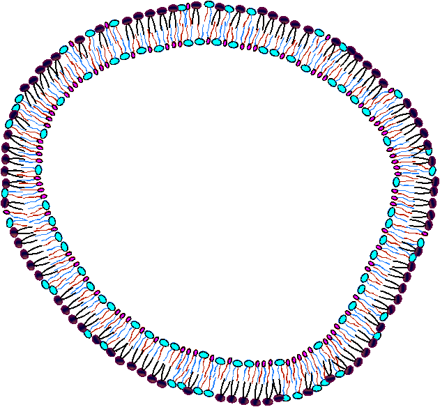
chromosome - elongate cellular structure composed of DNA and protein - they are the vehicles which carry DNA in cells
diploid (2n) - cellular condition where each chromosome type is represented by two homologous chromosomes
haploid (n) - cellular condition where each chromosome type is represented by only one chromosome
homologous chromosome - chromosome of the same size and shape which carry the same type of genes
chromatid - one of two duplicated chromosomes connected at the centromere
centromere - region of chromosome where microtubules attach during mitosis and meiosis
Chromosome structure
chromosome structure
composed of DNA and protein (histones) all tightly wrapped up in one package
duplicated chromosomes are connected by a centromere
2n=4 Example - an organism is 2n = 4.
Chromosomes 1 & 2 are homologous chromosomes
Chromosomes 3 & 4 are homologous chromosomes
Chromosomes 1 & 3 came from the mother
Chromosomes 2 & 4 came from the father
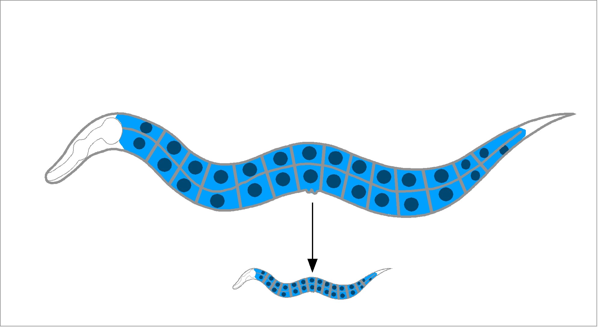
Typical Animal Life Cycle
animal life cycle
The Cell Cycle
cell cycle G1 - first gap
S - DNA synthesis (replication)
G2 - second gap
M - mitosis
mitosis - nuclear/chemical events resulting in two daughter nuclei which have identical genetic material to each other and to the mother cell
cytokinesis - division of the cytoplasm. This usually occurs with mitosis, but in some organisms this is not so
Mitosis in a Nutshell
The stages of the cell cycle can be broken down into six stages:
Interphase, Prophase, Metaphase, Anaphase, Telophase
Interphase
is the "resting" or non-mitotic portion of the cell cycle.
It is comprised of G1, S, and G2 stages of the cell cycle.
DNA is replicated during the S phase of Interphase
Prophase Prophase Prophase - the first stage of mitosis.
The chromosomes condense and become visible
The centrioles form and move toward opposite ends of the cell ("the poles")
The nuclear membrane dissolves
The mitotic spindle forms (from the centrioles in animal cells)
Spindle fibers from each centriole attach to each sister chromatid at the kinetochore
Compare Prophase to the Prophase I and to the Prophase II stages of mitosis.
Metaphase
The Centrioles complete their migration to the poles
The chromosomes line up in the middle of the cell ("the equator")
Compare Metaphase to the Metaphase I and to the Metaphase II stages of mitosis. Metaphase Metaphase
Anaphase Anaphase Anaphase
Spindles attached to kinetochores begin to shorten.
This exerts a force on the sister chromatids that pulls them apart.
Spindle fibers continue to shorten, pulling chromatids to opposite poles.
This ensures that each daughter cell gets identical sets of chromosomes
Compare Anaphase to the Anaphase I and to the Anaphase II stages of mitosis.
Telophase
The chromosomes decondense
The nuclear envelope forms
Cytokinesis reaches completion, creating two daughter cells
Compare Telophase to the Telophase I and to the Telophase II stages of mitosis. Telophase Telophase
Cytokinesis Divides the Cytoplasm
In animal cells, cytokinesis occurs by a process known as cleavage
First, a cleavage furrow appears
cleavage furrow = shallow groove near the location of the old metaphase plate
A contractile ring of actin microfilaments in association with myosin, a protein
Actin and myosin are also involved in muscle contraction and other movement functions
The contraction of a the dividing cell's ring of microfilaments is like the pulling of drawstrings
The cell is pinched in two
Cytokinesis in plant cells is different because plant cells have cell walls.
There is no cleavage furrow
During telophase, vesicles from the Golgi apparatus move along microtubules to the middle of the cell (where the cell plate was) and coalesce, producing the cell plate
Cell-wall construction materials are carried in the vesicles and are continually deposited until a complete cell wall forms between the two daughter cells
Simple Animal Cell Animal Cell Model Diagram Project Parts Structure Labeled Coloring and Plant Cell Organelles Cake

Simple Animal Cell Animal Cell Model Diagram Project Parts Structure Labeled Coloring and Plant Cell Organelles Cake
Simple Animal Cell Animal Cell Model Diagram Project Parts Structure Labeled Coloring and Plant Cell Organelles Cake
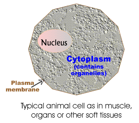
Simple Animal Cell Animal Cell Model Diagram Project Parts Structure Labeled Coloring and Plant Cell Organelles Cake

Simple Animal Cell Animal Cell Model Diagram Project Parts Structure Labeled Coloring and Plant Cell Organelles Cake

Simple Animal Cell Animal Cell Model Diagram Project Parts Structure Labeled Coloring and Plant Cell Organelles Cake

Simple Animal Cell Animal Cell Model Diagram Project Parts Structure Labeled Coloring and Plant Cell Organelles Cake

Simple Animal Cell Animal Cell Model Diagram Project Parts Structure Labeled Coloring and Plant Cell Organelles Cake
Simple Animal Cell Animal Cell Model Diagram Project Parts Structure Labeled Coloring and Plant Cell Organelles Cake

Simple Animal Cell Animal Cell Model Diagram Project Parts Structure Labeled Coloring and Plant Cell Organelles Cake

Simple Animal Cell Animal Cell Model Diagram Project Parts Structure Labeled Coloring and Plant Cell Organelles Cake
No comments:
Post a Comment